 Hội Nội Tiết – Đái Tháo Đường Miền Trung Việt Nam Hội Nội Tiết – Đái Tháo Đường Miền Trung Việt Nam
STUDY ON BONE MINERAL DENSITY, OSTEOPOROSIS BY DUAL ENERGY X – RAY ABSORPTION METRY (DEXA) IN TYPE 2 DIABETIC PATIENTS AT THE ENDOCRINE HOSPITAL OF THANH HOA PROVINCE
Tran Thua Nguyen1, Ha Khanh Du2, Le Thi Ngoc Uyen1
1. Hue Central Hospital.
2. Endocrine hospital of Thanh Hoa province
DOI: 10.47122/vjde.2022.53.5
ABSTRACT
Background: Secondary osteoporosis contributes to reduced quality of life, while increasing the risk of morbidity and mortality in type 2 diabetes patients. Diabetes and related complications can be detrimental to bone quality. Objective: Survey on bone mineral density, Osteoporosis by Dual energy X – ray absorptionmetry (DEXA) in type 2 diabetic patients at the Endocrine hospital of Thanh Hoa province; To evaluate the relation between bone mineral density, Osteoporosis and age, gender. Methods: A cross- sectional study on 1538 type 2 diabetes patients at the Endocrine hospital of Thanh Hoa province, from April 2019 to April 2021. Type 2 diabetes were diagnosed by Vietnam Ministry of Health Criteria. All patients were interviewed for getting information of age, gender. Bone mineral density were evaluated by Dual energy X – ray absorptionmetry (DEXA). Data were analysed by SPSS 13.0 software. Results: The mean value of bone mineral density at lumbar spine was 0.755 ± 0.151g/cm2; at femur neck was 0.774 ± 0.17 g/cm2. The percentage of decreased bone mineral density was 37%, in which: 32,74% for men and 40.84% for women. The percentage of osteoporosis was 13.46%, in which: 10.68% for men and 15.96% for women. The older the age, the lower the reduction of bone mineral density and the higher the rate of osteoporosis. Conclusion: It should be pay attention in the diagnosis and treatment of osteoporosis in patients with type 2 diabetes
Key words: bone mineral density, osteoporosis, type 2 diabetes
Main correspondence: Tran Thua Nguyen
Submission date: 19th Oct 2022
Revised date: 19th Nov 2022
Acceptance date: 19th Dec 2022
Email: [email protected]
Tel: + 84 234 903597695
- BACKGROUND
Osteoporosis is the most common disease in the elderly today, second only to cardiovascular disease. One-third of women and one-eighth of men over the age of 50 are at risk of osteoporosis. The disease causes many consequences, affecting quality of life and is the cause of increased mortality. Diagnosis of osteoporosis is based on bone density measurement [7], [11], [13].
In 2013, in the results announced by the “National Diabetes Prevention Project” implemented by the National Hospital of Endocrinology in 2012 on 11,000 people aged 30-69 in 6 regions including: Northern mountainous region, Delta region. The Red
River, Central Coast, Central Highlands, Southeast and Southwest regions showed a prevalence of diabetes of 5.7% [2].
Diabetes in general and type 2 diabetes in particular, if untreated or ineffectively treated, will cause many acute and chronic complications that are dangerous to life as well as quality of life, one of which is osteoporosis. Secondary osteoporosis in patients with type 2 diabetes contributes to a reduced quality of life and an increased risk of morbidity and mortality in patients.
Diabetes and related complications can be detrimental to bone quality [2].
The manifestations of osteoporosis are often discreet, difficult to detect early. When bone weight loss is about 30-40%, there are clinical signs such as: spinal pain, spinal deformity or fracture… Its consequences is to reduce mobility, reduce labor, reduce quality of life, and increase the economic burden on society. Therefore, proper assessment of bone health is an important step in the prevention of osteoporosis. If waiting for clinical manifestations to diagnose osteoporosis, it is too late. Currently, there are many advanced methods applied to evaluate bone density, in which the method of measuring double energy X-ray absorptiometry (Dual enegy X – ray
absorptionmetry – DEXA), at the column location. Lumbar spine and femoral neck are the most used and highly accurate methods. This is the gold standard for the diagnosis of osteoporosis.
We carry out this project with the aim of:
– Survey of bone density in patients with type 2 diabetes by DEXA method.
– Evaluate the relationship between bone density and osteoporosis with age and sex in patients with type 2 diabetes
2. RESEARCH SUBJECTS AND METHODS
2.1. Subjects, location, time of research
2.1.1. Study location and time:
– Location at Thanh Hoa Endocrine Hospital.
– Research period from April 2019 to April 2021.
2.1.2. Research subjects:
* Research subjects: Our study subjects are patients diagnosed with type 2 diabetes aged 40 to 60 years who are undergoing outpatient and inpatient treatment at Thanh Hoa Endocrine Hospital who satisfy the following criteria: :
* Diagnostic criteria for type 2 diabetes (according to the Ministry of Health of Vietnam, 2017) [3]
– Fasting venous blood glucose > 126 mg/dl (7 mmol/l) 8 hours after eating.
– Diagnosed with diabetes aged 40 years or older
– The patient consented to participate in the study.
*Exclusion criteria:
Excluded from the study when the patient had one of the following:
– Does not match the criteria for patient selection…
– Patients with diseases:
+ Endocrine: Multiple myeloma, Cushing’s syndrome, hyperparathyroidism,.
+ Acute disease: Blood infection, diabetic coma
– The patient gave up during the study.
2.2. Research Methods
2.2.1. Study Design: Cross-sectional Description
2.2.2. Research indicators
2.2.2.1. Clinical indicators: age, sex
2.2.2.2. Measure bone density: Measure bone density in the femoral neck and in the lumbar spine from L2 – L4 by DEXA method.
– Principle of measuring bone density by DEXA method
– Measurement technique: the patient lies on the table, the machine automatically moves to the position to be measured and automatically selects measurement parameters such as speed, measured dose. The technician controls the machine to operate according to the established procedure before the measurement to complete the measurement. It is required to measure 2 indexes: Bone mineral content (BMC) and bone mineral density (BMD). Measurement location: lumbar spine, femoral neck.
– Preparation: the patient is carefully instructed on the steps to take the measurement, the technician makes a sample first so that the
patient understands and does the necessary technique correctly.
Detector: Receives signals from 2 types of tissues: bone tissue and soft tissue.
– Analyze results
At the lumbar spine:
* Bone density (BMD) was measured in the L1- L4 region; Bone mineral content was measured in an anteroposterior cross section in
each region corresponding to the area of bone density measurement.
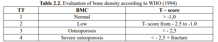
* The final result is calculated as the column average of the indicators in the measurement area. Bone density is displayed using the T-Score.
At the femoral neck:
* Bone density index was measured at the femoral neck, the great trochanter and the midpoint between the two above landmarks. The BMC index was measured in the anteroposterior section in each region corresponding to the bone density measurement region.
* The final result is the average of the indicators in the measurement area. Bone density is displayed using the T-Score.
+ Result: The computer automatically gives the T – Score: what is the value to compare the patient’s bone density with the bone density of young people, of the same sex, of the same race.
– Evaluation of bone density according to WHO (1994) [8].
2.2.3. Data processing: The obtained data were analyzed and calculated on the medical statistical software SPSS 13.0.
2.2.4. Control errors in research
– Uniform research medical record form for patients.
– Measurement of bone density by 1 person and on the same machine, trained at the department of osteopathy, Bach Mai hospital.
– Train technicians to measure blood pressure at the same mercury machine, measure height, weight (same weight), waist circumference, calculate BMI.
2.2.5. Ethics in research
– Approved by the Provincial Scientific Council as well as approved by the Leaders of Thanh Hoa Endocrine Hospital.
– Research results are informed to patients only to serve patients and improve the quality of medical examination and treatment.
– Patients and patients’ family members are clearly explained about the research objectives and content, voluntarily participate in the
research and have the right to withdraw from the study without explanation.
– The responses are only published as general information, not specifying the content of each individual’s reply.
– Research results are communicated to patients
3. RESEARCH RESULTS
3.1. Evaluation of bone density in patients with type 2 diabetes by DEXA method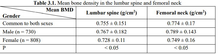
Comment: The BMD in the lumbar spine of men is 0.767 ± 0.182 (g/cm2) larger than the BMD in the lumbar spine of women (0.728 ± 0.11 (g/cm2)) (p < 0.05). The BMD at the femoral neck of men was 0.789 ± 0.143 (g/cm2) greater than the BMD of the femoral neck of women (0.749 ± 0.16 (g/cm2)) (p < 0.05)
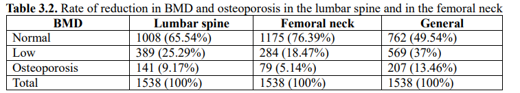
Comment: The overall rate of osteoporosis is 13.46%, the rate of reduction in overall bone density is 37%. The rate of bone density reduction in the lumbar spine is 25.29%, in the femoral neck is 18.47%. The rate of osteoporosis in the lumbar spine is 9.17%, in the femoral neck is 5.14%.
3.2. Evaluation of the relationship between BMD and osteoporosis with age, sex
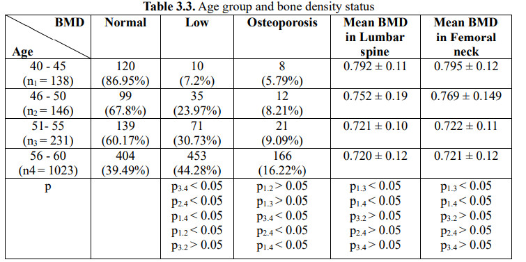
Comment: The rate of BMD reduction gradually increased by age group: 7.2%, 23.97%, 30.73%, 44.28% in the respective age groups: 40 – 45, 46 – 50, 51-55 , 56 – 60. The corresponding rate of osteoporosis is also: 5.79%, 8.21%, 9.09%, 16.22%.
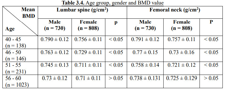
Comment: BMD in the lumbar spine of male aged 40 – 45; 46 – 50; 51 – 55 respectively: 0.790 ± 0.12 g/cm2; 0.763 ± 0.12 g/cm2; 0.745 ± 0.13 g/cm2 higher than female: 0.756 ± 0.11 g/cm2; 0.729 ± 0.11 g/cm2; 0.711 ± 0.11 g/cm2 of the same age group (p < 0.05), BMD in the femoral neck of male aged 40-45; 46 – 50; 51 – 55 respectively: 0.791 ± 0.12 g/cm2; 0.77 ± 0.15 g/cm2; 0.758 ± 0.14 g/cm2 higher than female: 0.757 ± 0.11 g/cm2; 0.73 ± 0.16 g/cm2; 0.721 ± 0.12 g/cm2 of the same age group (p < 0.05).
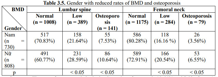
Comment: The rate of osteoporosis in the lumbar spine of women is 10.64% and 7.53 of men (p < 0.05). The rate of osteoporosis in the femoral neck of women is 6.55% and 3.56% of men (p < 0.05).
4. DISCUSSION
4.1. Bone density in patients with type 2 diabetes
4.1.1. Mean bone density in the femoral neck and lumbar spine
Through the study of 1538 subjects with type 2 diabetes, we found that the mean BMD in the lumbar spine was 0.755 ± 0.151 g/cm2
(In which the mean BMD in the lumbar spine of the male patient was 0.767 ± 0.182 g/cm2, the female patient is 0.728 ± 0.11 g/cm2); mean. BMD at femoral neck is 0.774 ± 0.17 g/cm2 ;(where mean BMD at femoral neck of male patient is 0.789 ± 0.143 g/cm2, female patient is 0.749 ± 0.16 g/cm2) (Table 3.1). Our results are lower than those of M.Yamamoto, T.Yamaguchi studied in Japan in 2007 [18] in patients with type 2 diabetes who had an average BMD at the femoral neck of 0.816 g/cm2, in the lumbar spine was 0.716 g/cm2, according to research by Ngo Mai Xuan, Tran Duc Tho in Hanoi [17]: mean BMD in femoral neck was 0.83 ± 0.11 g/cm2 and in lumbar spine was 0.83 ± 0.11 g/cm2 0.91 ± 0.11 g/cm2.
Meanwhile, our results are similar to the study of Dao Thi Dua in Hue: Mean BMD in femoral neck is 0.787 ± 0.132 g/cm2 and in lumbar spine is 0.75 ± 0.171 g/cm2 compared with the control group without diabetes: Mean BMD at femoral neck was 0.951 ± 0.154 g/cm2 and Mean BMD in lumbar spine was 0.932 ± 0.114 g/cm2 (p < 0.05) [4].
Dian L Chau and Steven V, when comparing BMD in patients with type 2 diabetes in Asia and Europe, noted that BMD in patients with
type 2 diabetes in Asia both decreased.
4.1.2. Rate of reduction in bone density, osteoporosis.
In our study, the rate of BMD reduction in patients with type 2 diabetes is 37% (in which, BMD reduction in femoral neck is 18.47%,
BMD reduction in lumbar spine is 25.29%), osteoporosis is 13.46% (in which, osteoporosis in the femoral neck is 5.14%, osteoporosis in the lumbar spine is 9.17%, (Table 3.2). Our results are lower than the study of Bach Thi Hoai Duong (2019) the rate of BMD reduction was 54.3% (BMD reduction in lumbar spine 24.3%, in femoral neck 38.6%).
Overall osteoporosis was 45.7% (in which, osteoporosis in lumbar spine 35.7%, femoral neck 32.9%) [6].
Dao Thi Dua (Hue) the rate of osteoporosis in the femoral neck of patients with type 2 diabetes was 36.37%, the reduction in BMD
was 40%; Osteoporosis in the spine is 50%, the reduction in BMD in the spine was 35% [4], [5].
Nguyen Thi Nguyen Trang, Nguyen Hai Thuy had up to 31.91% of patients with type 2 diabetes had osteoporosis. In the study of Nguyen Dinh Duyet, up to 84% of patients with type 2 diabetes had osteoporosis [16].
Ngo Mai Xuan, Tran Duc Tho, Do Trung Quan in Hanoi, the rate of osteoporosis in the femoral neck of patients with type 2 diabetes
was 8% and osteoporosis in the spine was 10% [18].
In the NC by Shivank Prakash et al: the overall rate of osteoporosis was 43.8% (osteoporosis in the spine 39.6%), the reduction in BMD was 26.05% [10].
Lin Xu (China): 29-31% of patients with type 2 diabetes had osteoporosis. Yhusao SI (China), in 2017: the rate of osteoporosis in patients with type 2 diabetes was 37.8%. The study of Balram Sharma et al in India (2015- 2016): the rate of osteoporosis in patients with type 2 diabetes was 35.5% (in the lumbar spine it was 33.5%, in the femoral neck it was 13.5%) [1].
The rate of reduction of BMD and osteoporosis in our lumbar spine and femoral neck is lower than that of some domestic and foreign authors, possibly because the age of the studies > 60 accounted for the majority and at the same time, our sample size is many times
larger than that of other studies.
4.2. Relationship between BMD and osteoporosis with age, sex
4.2.1. Relationship between age and bone density and osteoporosis
In our study: mean BMD in the femoral neck, in the lumbar spine gradually decreased with age leading to an increase in the rate of BMD reduction and osteoporosis with age. BMD reduction rate is 7.2%, osteoporosis rate 5.79% in age group (40 – 45) is lowest and highest in age group (56 – 60) 44.28% decrease in BMD, 16.22 % osteoporosis (p < 0.05) (Table 3.3).
Our results are similar to those of Bach Thi Hoai Duong [6], Nguyen Nguyen Trang, Nguyen Hai Thuy: the rate of BMD reduction in patients ≥ 60 years old was 48.94%, osteoporosis 27.66% (oppositely) In the group of patients under 45 years old, the rate of BMD reduction was 2.13%) [15].
Nguyen Hai Thuy (2008) recorded that 20- 30% over 60 years old had symptoms of osteoporosis in the spine without even knowing it. According to Nguyen Cong Hoa (2008), the rate of osteoporosis among people over 45 years old in Go Vap District, Ho Chi Minh City was 30.4%. According to Nguyen Thi Anh Thu (2006), 19.6% of women in their 50s. – 70 had osteoporosis, this rate increased to 58.8% in people over 70 years old. The rate in men also increased similarly.
Bach Thi Hoai Duong (2019): patients with type 2 diabetes (age < 60): the rate of bone loss in the lumbar spine 20.8%, osteoporosis in the femoral neck 16.7% was significantly lower than that of group (age > 60 years) osteoporosis in the lumbar spine 50%, osteoporosis in the femoral neck 43.2% [6]. Le Thi Hue in 113 elderly patients > 50 recorded that the rate of osteoporosis in the 60-79 group was 69.1%, the group 80 years old was 80.8%, the older the age, the higher the osteoporosis rate.
In the study of Shivank Prakash and CS: Age (40 – 45), reduced BMD 16.6%, osteoporosis 16.7%; age (46 – 50) reduced BMD 33.3%, osteoporosis 11.1%; age (51 – 55) reduced BMD 26.7%, osteoporosis 46.7%; age (56 – 60) reduced BMD 36.6%, osteoporosis 45.4%; age > 60 osteoporosis 51% [10]. Osteoporosis is caused by the aging process of osteoblasts, causing an imbalance between bone resorption and bone formation, which increases with age. Therefore, age is a risk factor for osteoporosis in patients with type 2 diabetes.
4.2.2. Relationship between gender and bone density and osteoporosis.
In our study, the rate of BMD reduction in female lumbar spine is 25.89% higher than 21.64% in men (p < 0.05). The rate of osteoporosis of lumbar spine in women 10 ,64% is higher than 7.53% in men and the rate of reduction of BMD in female femoral neck is 20.54% which is also higher than 16.16% in men. So is osteoporosis in the femoral neck (6.55% > 3.56% in men) (p < 0.05) (Table 3.5). When comparing the BMD in the femoral neck or the lumbar spine between the two sexes, it is found that at the age of 40 – 55 BMD in the two above positions in men were all large in women (p < 0.05). But from the age of 56 onwards, the average BMD in the lumbar spine and femoral neck in men and women aged 56 – 60 is not statistically significant (p > 0.05) (Table 3.4).
According to Nguyen Thi Anh Thu (2006), 19.6% of women aged 50 – 70 had osteoporosis, this rate increases to 58.8% in people over 70 years old. The rate in men also increased similarly.
Ngo Thi Thu Trang (2009 – 2012): the rate of reduction of lumbar spine BMD in female patients with type 2 diabetes was 0.738 ± 0.164 g/cm2 lower than 0.903 ± 0.187 g/cm2 in male patients with type 2 diabetes and the rate of osteoporosis at CL 62.5% was 36.8% higher than that of men, the rate of reduction of BMD in femoral neck of female patients with type 2 diabetes 0.695 ± 0.161g/cm2 was lower than 0.782 ± 0.203 g/cm2 in male patients Type 2 diabetes and osteoporosis in the femoral neck 38.5% were 18.4% higher than in men (p < 0.05) [16]. Le Thanh Toan: BMD in the femoral neck of female patients was 0.62 ± 0.15 g/cm2 lower than that of male patients by 0.76 ± 0.16 g/cm2; The rate of osteoporosis in male patients (30.2%) was lower than in female patients (64.8%). Le Thi My Linh: the rate of osteoporosis in male patients (30.2%) compared with female patients (41.8%) [14].
Research by Nguyen Thi Phuong Thuy recorded the rate of osteoporosis in elderly women 66.7%, older men 15.2% [12].
Women are four times more likely than men to have primary osteoporosis because of their lower bone mass and faster bone loss than men as a result of the rapid decline in ovarian function after menopause [9].
5. CONCLUSION
Through the study of 1538 subjects with type 2 diabetes aged 40-60 years old at Thanh Hoa Endocrinology Hospital, we drew some conclusions as follows:
5.1. About bone density
– The average bone density in the lumbar spine is 0.755 ± 0.151 g/cm2
– The average bone density at the femoral neck was 0.774 ± 0.17 g/cm2
– The rate of bone density reduction is 37% (in lumbar spine: 25.29%, femoral neck is 18.47%). In which (Male reduced bone density: 32.74%, Female bone density decreased 40.84%).
– Osteoporosis rate 13.46% (lumbar spine is 9.17%, femoral neck is 5.14%).
5.2. On the relationship between bone density and osteoporosis with age, sex
– The rate of osteoporosis in men is 10.68%, in women is 15.96%.
– Women have lower average bone density in the lumbar spine and in the femoral neck than in men; the rate of bone density loss and osteoporosis is higher in women than in men. With age, bone density decreases and the rate of osteoporosis increases.
REFERENCES
1. Balram Sharma, Hema Singh, Praveen Chodhary, Sanjay Saran, Sandeep Kumar Mathur (2017), “Osteoporosis in otherwise healthy patients with type 2 diabetes: A prospective gender based comparative study”, Journal of Clinical Densitometry, 21(4): 535-539.
2. Ta Van Binh (2003), “Diabetes epidemiology – Risk factors and issues related to diabetes management in the inner city of 4 big cities”, Publishing House Medicine, Hanoi
3. Ministry of Health (2017), “Guidelines for the diagnosis and treatment of type 2 diabetes”, Issued under Decision No. 3319/QD-BYT.
4. Dao Thi Dua, Nguyen Minh Quang, Hoang Khanh Hang (2009), “Surveying the relationship between bone density and blood calcium and phosphorus levels and some risk factors for osteoporosis in diabetic patients type 2″, Journal of Internal Medicine No. 4/2009, pp. 293 – 300.
5. Dao Thi Dua (2010), “Research on osteoporosis in patients with type 2 diabetes”, Hue University Hospital of Medicine and Pharmacy, Practical medicine. December 2010.
6. Bach Thi Hoai Duong, Nguyen Dinh Toan (2020), “Study on the prevalence of osteoporosis and some factors related to osteoporosis in patients with type 2 diabetes”, Journal of Endocrinology & Diabetes, number 39, p. 66 – 71.
7. Pham Minh Duc (1996), “Metabolism and regulation of calcium phosphate metabolism”, Physiology, Volume 1, Medical Publishing House, p. 113-129.
8. Nguyen Thi Ngoc Lan (2018), “Osteoporosis”, Internal Pathology, Volume II, Hanoi Medical Publishing House, p. 205 – 210.
9. Le Thi My Linh (2010), Research on the degree of osteoporosis in patients with type 2 diabetes at Cho Ray Hospital”, Master’s Thesis of Medicine, University of Medicine and Pharmacy, HCMC. Ho Chi Minh.
10. Shivank Prakash, Ravi. S. Jatti, Shridhar C. Ghagane, S.M. Jali and M.V. Jali (2012), “Prevalence of Osteoporosis in Type 2 Diabetes Mellitus Patients Using Dual Energy X-Ray Absorptiometry (DEXA) Scan”. International Journal of Osteoporosis and Metabolic Disorders, 10:10-16.
11. Tran Duc Tho (1999), “Osteoporosis in the elderly”, Hanoi Medical Publishing House.
12. Nguyen Thi Phuong Thuy (2012), Research on osteoporosis in elderly patients with type 2 diabetes”, Thesis of Master of Medicine. Hanoi Medical University.
13. Vu Thi Thanh Thuy, Cao Thi Nhi (1995), “Methods to treat osteoporosis”, Journal of Internal Medicine No. 1, p. 15 – 17.
14. Le Thanh Toan et al. (2012), “Study on bone density by dexa method in diabetic patients at Cho Ray Hospital”, Medical Journal of Ho Chi Minh City. Ho Chi Minh, volume 16, p. 348-353.
15. Nguyen Nguyen Trang, Nguyen Hai Thuy (2010), “Survey on bone density in patients with type 2 diabetes”, Journal of Internal Medicine No. 4/2010, p. 301-312
16. Ngo Thi Thu Trang, Nguyen Thi Phi Nga (2013), “Study on bone density, osteoporosis rate by dual-energy X-ray absorptiometry (DEXA) in female patients with type 2 diabetes”, Journal of Military Medicine – Pharmacology, No. 2, p. 47 – 53.
17. Ngo Thi Mai Xuan (2006), Evaluation of bone density in female patients with type 2 diabetes mellitus by DEXA method, Level 2 specialized thesis.
18. Ngo Mai Xuan, Tran Duc Tho, Do Trung Quan (2009), “Review of bone density in female patients with type 2 diabetes and related factors”, Practical medicine, 673, p. 315-324